

Cross-section esophagus of the dog. Medical teaching equipment and biological tissue slide. Educational material for the study and treatment of doggy. Colouring with hematoxylin and eosin.
Коллекция по умолчанию
Коллекция по умолчанию
Создать новую

Histopathology of pneumonia, light micrograph, photo under microscope. Cellulose aspiration pneumonia
Коллекция по умолчанию
Коллекция по умолчанию
Создать новую

Blast cells in leukemia.blood smear is often used as a follow-up test to abnormal results on a complete blood count (CBC) to evaluate the different types of blood cells.
Коллекция по умолчанию
Коллекция по умолчанию
Создать новую

Sperm and egg cell on scientific background. 3d illustration
Коллекция по умолчанию
Коллекция по умолчанию
Создать новую

Abnormal neutrophil in pleural fluid smear.
Коллекция по умолчанию
Коллекция по умолчанию
Создать новую

Prostate cancer, light micrograph, photo under microscope
Коллекция по умолчанию
Коллекция по умолчанию
Создать новую

Blood picture of acute myeloid leukemia
Коллекция по умолчанию
Коллекция по умолчанию
Создать новую

Malaria parasite P. vivax
Коллекция по умолчанию
Коллекция по умолчанию
Создать новую

Caseous necrosis of lymphatic node, light micrograph, photo under microscope
Коллекция по умолчанию
Коллекция по умолчанию
Создать новую

some strange molecules enlarged under a microscope
Коллекция по умолчанию
Коллекция по умолчанию
Создать новую

Abstract creative marbling pattern for fabric, design background texture
Коллекция по умолчанию
Коллекция по умолчанию
Создать новую

Basal cell carcinoma, skin cancer, light micrograph, photo under microscope
Коллекция по умолчанию
Коллекция по умолчанию
Создать новую

Squamous cell carcinoma, light micrograph, photo under microscope
Коллекция по умолчанию
Коллекция по умолчанию
Создать новую

Basal cell carcinoma, skin cancer, light micrograph, photo under microscope
Коллекция по умолчанию
Коллекция по умолчанию
Создать новую

Renal tuberculosis, light micrograph, photo under microscope
Коллекция по умолчанию
Коллекция по умолчанию
Создать новую

bacteria with epithelium cell in gram stain method.
Коллекция по умолчанию
Коллекция по умолчанию
Создать новую

Renal cell carcinoma, light micrograph, photo under microscope
Коллекция по умолчанию
Коллекция по умолчанию
Создать новую

Ovarian follicular cyst, light micrograph, photo under microscope
Коллекция по умолчанию
Коллекция по умолчанию
Создать новую

Chronic bronchitis, photo under microscope, light micrograph
Коллекция по умолчанию
Коллекция по умолчанию
Создать новую

Acute myocardial infarction, histology of heart tissue, light micrograph. Area of infarct is paler than than the relatively viable area of heart muscle
Коллекция по умолчанию
Коллекция по умолчанию
Создать новую

Villous colon adenocarcinoma, light micrograph, photo under microscope
Коллекция по умолчанию
Коллекция по умолчанию
Создать новую

Purple water drink bubbles abstract background
Коллекция по умолчанию
Коллекция по умолчанию
Создать новую

Histopathology of alcoholic hepatitis, light micrograph, photo under microscope
Коллекция по умолчанию
Коллекция по умолчанию
Создать новую

A blood smear is often used as a follow-up test to abnormal results on a complete blood count (CBC) to evaluate the different types of blood cells.
Коллекция по умолчанию
Коллекция по умолчанию
Создать новую

Chronic pyelonephritis, light micrograph, photo under microscope
Коллекция по умолчанию
Коллекция по умолчанию
Создать новую

Histopathology of lung emphysema, light micrograph, photo under microscope showing enlargement of air spaces in lung tissue and destruction of alveolar septa
Коллекция по умолчанию
Коллекция по умолчанию
Создать новую

Ovarian cancer, light micrograph, photo under microscope. Photograph shows a fragment of a cancerous tumor in the female ovary. Selective focus
Коллекция по умолчанию
Коллекция по умолчанию
Создать новую

Uterine cancer, light micrograph, photo under microscope
Коллекция по умолчанию
Коллекция по умолчанию
Создать новую

Scenic background from spots and stains of oil paint in purple tones
Коллекция по умолчанию
Коллекция по умолчанию
Создать новую

Colon adenocarcinoma, light micrograph, photo under microscope
Коллекция по умолчанию
Коллекция по умолчанию
Создать новую

Breast ductal carcinoma, light micrograph, photo under microscope
Коллекция по умолчанию
Коллекция по умолчанию
Создать новую

Serpentine - rock of serpentine group minerals. Background
Коллекция по умолчанию
Коллекция по умолчанию
Создать новую

Tissue of Small intestine (Duodenum), Large intestine Human and Stomach Human under the microscope in Lab.
Коллекция по умолчанию
Коллекция по умолчанию
Создать новую

Histopathology of hypertensive renal disease, light micrograph, photo under microscope
Коллекция по умолчанию
Коллекция по умолчанию
Создать новую

Acute promyelocytic leukemia cells or APL, analyze by microscope, original magnification 1000x
Коллекция по умолчанию
Коллекция по умолчанию
Создать новую

Well-differentiated intestinal adenocarcinoma, light micrograph, photo under microscope
Коллекция по умолчанию
Коллекция по умолчанию
Создать новую

multinucleated giant
Коллекция по умолчанию
Коллекция по умолчанию
Создать новую

Gram staining, also called Gram's method, is a method of differentiating bacterial species into two large groups (Gram-positive and Gram-negative).
Коллекция по умолчанию
Коллекция по умолчанию
Создать новую

Fibrinoid necrosis of the vessel wall, light micrograph, photo under microscope
Коллекция по умолчанию
Коллекция по умолчанию
Создать новую

Histopathology of biliary liver cirrhosis, light micrograph, photo under microscope
Коллекция по умолчанию
Коллекция по умолчанию
Создать новую

blood films for Malaria parasite.showing pink cells malaria pigment.medical science background concept.
Коллекция по умолчанию
Коллекция по умолчанию
Создать новую

Chronic nephritis, light micrograph, photo under microscope. High magnification
Коллекция по умолчанию
Коллекция по умолчанию
Создать новую

Kidney cancer, light micrograph, photo under microscope
Коллекция по умолчанию
Коллекция по умолчанию
Создать новую

Bladder cat- cell nature background. Abstract- photo macro sections with high magnification with light microscope
Коллекция по умолчанию
Коллекция по умолчанию
Создать новую

Structure of Tissue of Spleen Human, Liver Human and Kidney Human under the microscope in Lab.
Коллекция по умолчанию
Коллекция по умолчанию
Создать новую

Squamous cell carcinoma, light micrograph, photo under microscope
Коллекция по умолчанию
Коллекция по умолчанию
Создать новую

Histopathology of interstitial nephritis, light micrograph, photo under microscope
Коллекция по умолчанию
Коллекция по умолчанию
Создать новую

Human blood smear showing a lymphocyte. The small bluish dots among the red blood cells are platelets.
Коллекция по умолчанию
Коллекция по умолчанию
Создать новую

Gram staining, also called Gram's method, is a method of differentiating bacterial species into two large groups (Gram-positive and Gram-negative).bacteria cells on pink background.
Коллекция по умолчанию
Коллекция по умолчанию
Создать новую

White blood cells in in blood smear, analyze by microscope
Коллекция по умолчанию
Коллекция по умолчанию
Создать новую

Watercolor hand drawn bubbles colorful soap froth
Коллекция по умолчанию
Коллекция по умолчанию
Создать новую

Histopathology of lung emphysema, light micrograph, photo under microscope showing enlargement of air spaces in lung tissue and destruction of alveolar septa
Коллекция по умолчанию
Коллекция по умолчанию
Создать новую

Tissue of Stomach Human under the microscope in Lab.
Коллекция по умолчанию
Коллекция по умолчанию
Создать новую

Abstract colored background from spilled paints closeup.
Коллекция по умолчанию
Коллекция по умолчанию
Создать новую

Bacteria cells infection in epithelial cells.
Коллекция по умолчанию
Коллекция по умолчанию
Создать новую

Characteristics Tissue of Olfactory epithelium Human under the microscope in Lab.
Коллекция по умолчанию
Коллекция по умолчанию
Создать новую

Histopathology of intestinal adenoma, light micrograph, photo under microscope
Коллекция по умолчанию
Коллекция по умолчанию
Создать новую

Acute hemorrhagic pancreatitis, light micrograph, hematoxylin and eosin staining
Коллекция по умолчанию
Коллекция по умолчанию
Создать новую

Histopathology of interstitial nephritis, light micrograph, photo under microscope
Коллекция по умолчанию
Коллекция по умолчанию
Создать новую

Acute myocardial infarction, histology of heart tissue, light micrograph. Area of infarct is paler than than the relatively viable area of heart muscle
Коллекция по умолчанию
Коллекция по умолчанию
Создать новую

Histology of human kidney, light micrograph showing nephron. Microscopy, hematoxylin and eosin staining
Коллекция по умолчанию
Коллекция по умолчанию
Создать новую

mesothelial cell in pleural fluid
Коллекция по умолчанию
Коллекция по умолчанию
Создать новую

Host cells with spores (mold) are inside wood under the microscope for education.
Коллекция по умолчанию
Коллекция по умолчанию
Создать новую

Light micrograph of chlorophyte green algae Volvox
Коллекция по умолчанию
Коллекция по умолчанию
Создать новую

Breast fibroadenosis, light micrograph, photo under microscope. Common benign hyperplastic process involving breast glands
Коллекция по умолчанию
Коллекция по умолчанию
Создать новую

Breast ductal carcinoma, light micrograph, photo under microscope
Коллекция по умолчанию
Коллекция по умолчанию
Создать новую

Benign prostatic hyperplasia. Micrograph shows dilated glands, papillary projections inside the lumen of the glands, cystic dilatation with accumulation of secretory material. Photo under microscope
Коллекция по умолчанию
Коллекция по умолчанию
Создать новую

Bubble Grunge mixed Hand Made
Коллекция по умолчанию
Коллекция по умолчанию
Создать новую

Anatomy and Histological Bone, Elastic cartilage human and Joint of human foetus under the microscope for education.
Коллекция по умолчанию
Коллекция по умолчанию
Создать новую

multinucleated giant
Коллекция по умолчанию
Коллекция по умолчанию
Создать новую

Gout tophus, photomicrograph showing amorphous, eosinophilic material with a foreign-body giant cell reaction and inflammation.
Коллекция по умолчанию
Коллекция по умолчанию
Создать новую

Gout tophus, photomicrograph showing amorphous, eosinophilic material with a foreign-body giant cell reaction and inflammation.
Коллекция по умолчанию
Коллекция по умолчанию
Создать новую

Blood picture of acute myeloid leukemia (AML)
Коллекция по умолчанию
Коллекция по умолчанию
Создать новую

White wooden texture with violet grass and moss. White bark natural beautiful background with purple veins.
Коллекция по умолчанию
Коллекция по умолчанию
Создать новую

Histopathology of interstitial nephritis, light micrograph, photo under microscope
Коллекция по умолчанию
Коллекция по умолчанию
Создать новую

Breast fibroadenosis, light micrograph, photo under microscope. Common benign hyperplastic process involving breast glands
Коллекция по умолчанию
Коллекция по умолчанию
Создать новую

Pike kidney in cross section 100x
Коллекция по умолчанию
Коллекция по умолчанию
Создать новую

Contracted kidney, light micrograph, photo under microscope
Коллекция по умолчанию
Коллекция по умолчанию
Создать новую

Uterine cancer, light micrograph, photo under microscope
Коллекция по умолчанию
Коллекция по умолчанию
Создать новую

Histopathology of nasal polyps, light micrograph, photo under microscope
Коллекция по умолчанию
Коллекция по умолчанию
Создать новую

multinucleated giant
Коллекция по умолчанию
Коллекция по умолчанию
Создать новую

Human blood smear showing a lymphocyte. The small bluish dots among the red blood cells are platelets.
Коллекция по умолчанию
Коллекция по умолчанию
Создать новую

Ink marble texture. Ebru handmade wave background. Kraft paper surface. Unique art illustration. Liquid marbling texture.
Коллекция по умолчанию
Коллекция по умолчанию
Создать новую

3D Illustration red blood cells in vein. Red blood cells flow in vessel. medical human health-care concept
Коллекция по умолчанию
Коллекция по умолчанию
Создать новую

pap smear, NILM (class II non specific chronic inflammation) under a microscope view for education histology. Human tissue.
Коллекция по умолчанию
Коллекция по умолчанию
Создать новую

Histopathology of acute nephritis, light micrograph, photo under microscope
Коллекция по умолчанию
Коллекция по умолчанию
Создать новую

Histopathology of hypertensive renal disease, light micrograph, photo under microscope
Коллекция по умолчанию
Коллекция по умолчанию
Создать новую
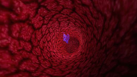
3d illustration of Virus Through A Vein Or Artery
Коллекция по умолчанию
Коллекция по умолчанию
Создать новую

Коллекция по умолчанию
Коллекция по умолчанию
Создать новую

plasmodium vivax early schizont state
Коллекция по умолчанию
Коллекция по умолчанию
Создать новую

A cross section of ciliated epithelium under the microscope.
Коллекция по умолчанию
Коллекция по умолчанию
Создать новую

Parasitic plant fungus Puccinia microscope slide.
Коллекция по умолчанию
Коллекция по умолчанию
Создать новую

Shampoo bubbles- dish soap. Drops- liquid foam. Fluid aqua- abstract pattern nature. Background- cleansing wash
Коллекция по умолчанию
Коллекция по умолчанию
Создать новую

Diffuse sclerosing glomerulonephritis, light micrograph, photo under microscope. High magnification
Коллекция по умолчанию
Коллекция по умолчанию
Создать новую

bursts of multicolored paints and mixed together white paper. Drawn splashes of paints of different colors on white surface. Blue, yellow, red, green color. Spreading spraying flow leak stream mixing
Коллекция по умолчанию
Коллекция по умолчанию
Создать новую

Chronic nephritis, light micrograph, photo under microscope
Коллекция по умолчанию
Коллекция по умолчанию
Создать новую

Villous colon adenocarcinoma, light micrograph, photo under microscope
Коллекция по умолчанию
Коллекция по умолчанию
Создать новую

Structure of Tissue of Spleen Human, Liver Human and Kidney Human under the microscope in Lab.
Коллекция по умолчанию
Коллекция по умолчанию
Создать новую

Breast cancer, light micrograph, photo under microscope
Коллекция по умолчанию
Коллекция по умолчанию
Создать новую

Leukemia cells in blood smear
Коллекция по умолчанию
Коллекция по умолчанию
Создать новую
Legion-Media
Создайте свои проекты на основе качественных стоковых фотографий и видео.


Copyright © Legion-Media.